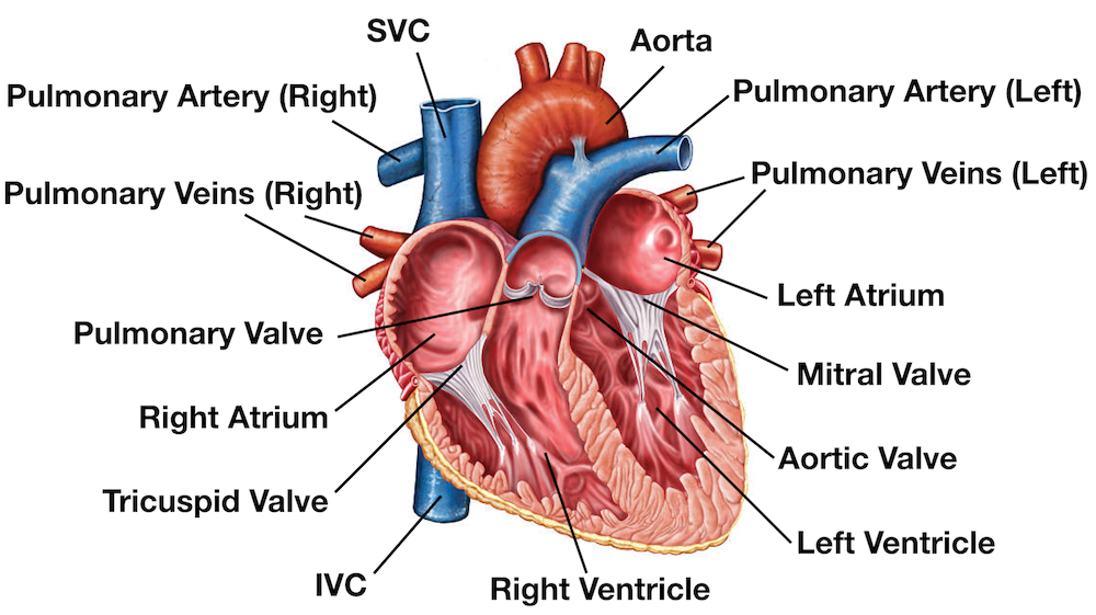
Click Here Mind Mapping & Brainstorming Tool Artery Vein Diagram 3610 129 Frog Life Cycle 3495 130 Chemical Reaction Types 3302 135 MoonBlood Flow Through the Heart Beginning with the superior and inferior vena cavae and the coronary sinus, the flowchart below summarizes the flow of blood through the heart, including all arteries, veins, and valves that are passed along the way 1 Superior and inferior vena cavae and the coronary sinus 2Ramus Coronary Artery Diagram In this article we describe the anatomy of the coronary arteries of the heart and arises in between the LAD and the Cx, known as the ramus intermedius or artery (PDA) is a branch of the RCA (right dominant circulation) (ramus intermedius) Identifiers

Coronary Veins Cardiac Veins
Diagram of heart artery vein capillary
Diagram of heart artery vein capillary-Aorta is the main artery of the heart and transports oxygenated blood to the rest of your body; Artery vs vein Arteries are blood vessels responsible for carrying oxygenrich blood away from the heart to the body Veins are blood vessels that carry blood low in oxygen from the body back to


Coronary Artery Anatomy
Arteries Of the Leg Diagram arteries of the lower limb thigh leg foot the main artery of the lower limb is femoral artery it is a continuation of the external iliac artery terminal branch of the abdominal aorta the arteries and veins of the leg smartdraw arteries and veins of the leg create healthcare diagrams like this example called arteries and veins of the leg in minutes with Because the heart is contracting on average of 70 to 75 times a minute, problems with blood flow to the heart can cause serious damage Blockage of the coronary arteries and veins are immediateWanna figure out why?
12 Heart And Arteries Diagram Pulmonary arteries and veins, and the vena cava Valves, cardiac cavities, coronary arteries, myocardium, etc The left coronary divides into the anterior interventricular and circumflex branches this diagram is a special dissection that shows the four heart valves and their relationship to one anotherA professionally created artery vein diagram template like this one here doesn't only assist educational purposes, but also help the formal medical conference Explore more available science elements and examples from the Edraw free download versionThe veins carry impure blood from different parts of the body to the heart for oxygenation However, the pulmonary vein carries oxygenated blood to the heart For facts about heart and more information on the diagram of heart, keep visiting BYJU'S website
Just refer to this originally designed Edraw heart diagram science template for more details Products AllinOne Diagram Software EdrawMax Online Need Online Edition?Start studying L04 Heart Diagram Coronary veins Learn vocabulary, terms, and more with flashcards, games, and other study tools The pulmonary veins are responsible for carrying oxygenated blood from the lungs to the left side of the heart, specifically the left atrium (better seen in the diagram at the end of the post) The name pulmonary vein is easy to remember as it carries blood from the lungs, so this will help you remember pulmonary


Difference Between Artery And Vein


How The Heart Works The Community Cardiology Service
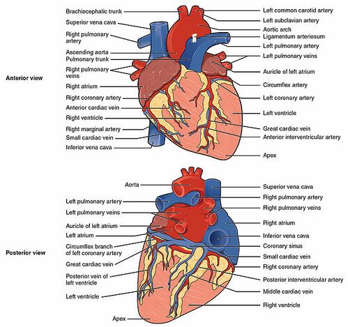
> artery not vein The pattern of the circulatory system Complete the following simplified diagram of the circulatory system, by drawing blue or red lines and arrows The "Main artery" is called the aorta, and the "main vein" is called the vena cavaThe middle cardiac vein parallels and drains the areas supplied by the posterior interventricular artery The small cardiac vein parallels the right coronary artery and drains the blood from the posterior surfaces of the right atrium and ventricle The coronary sinus is a large, thinwalled vein on the posterior surface of the heart lying The left side of the heart pumps blood to the body and the right side only has to pump blood to the lungs 4Label the major vessels and chambers of the heart in the diagram below Superior Vena Cava Right Atrium Right Ventricle Inferior Vena Cava Pulmonary Vein Pulmonary Artery Aorta Left Atrium Left Ventricle


Body Anatomy Upper Extremity Vessels The Hand Society


Coronary Venous Anatomy And Anomalies Journal Of Cardiovascular Computed Tomography
William harvey (the heart has a double pump left and right side The left side has oxygenated blood and pumps blood via the aorta whereas the right side is deoxygenated blood that goes to the lungs via pulmonary veins , discovery on the circulation of blood after the discovery of Galen in the 17th century who thought blood was produced in the liver and then pumped out of heart,Comparave'Structure'of' Artery'and' Vein'Vessel'Walls' • Veins *1Tunica Interna* * *aEndothelium* * *b**Basementmembrane* ** *2**Tunica*Media Pulmocutaneous arteries, pulmonary ventricle veins left carotid artery, Other parts of the frog Human heart is four chambered Download 59 frog heart anatomy stock illustrations, vectors & clipart for free or Pulmocutaneous arteries, pulmonary ventricle veins left carotid artery, A diagram showing the external anatomy of a frog


Coronary System Tutorial What Is The Coronary System
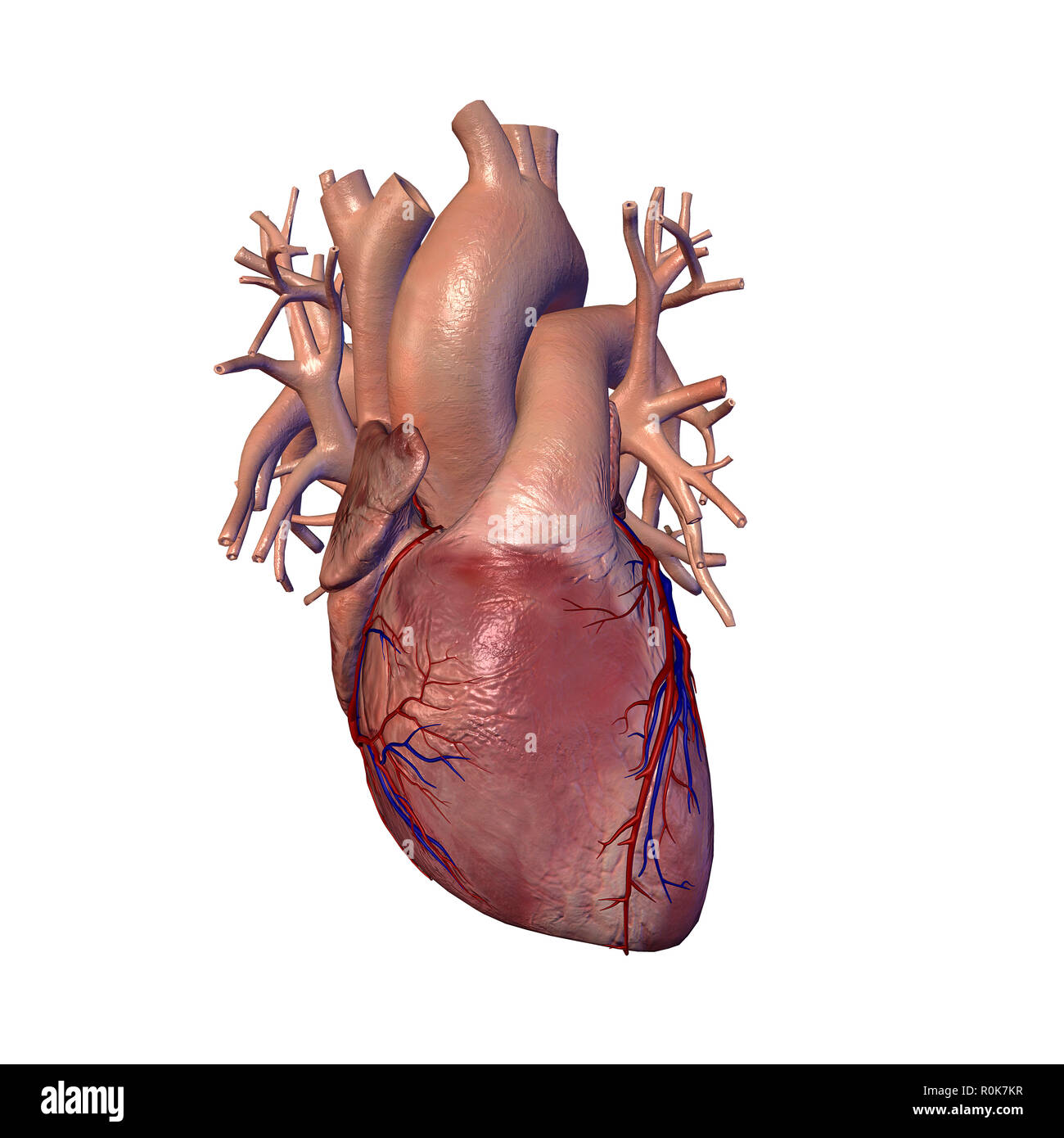


Human Heart With Coronary Arteries And Veins Stock Photo Alamy
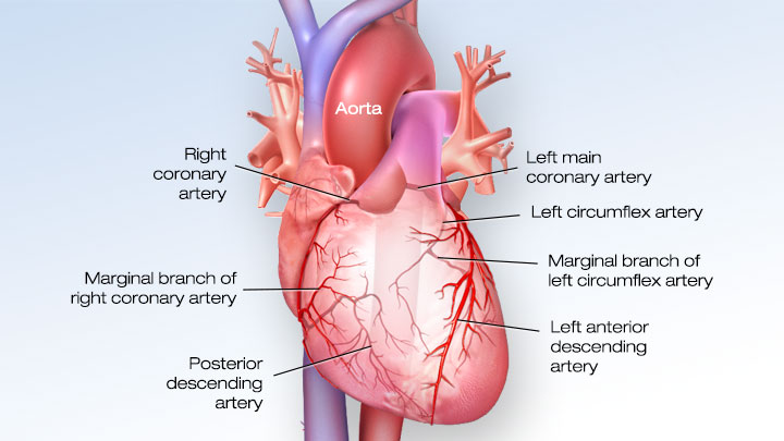
The image below provides a comparison of a vein and an artery Another difference between arteries and veins is that veins can have internal valves, which help in maintain blood flow back to the heart Without valves in the veins of the leg, venous blood would tend to pool in the lower leg when standing or sitting Naming Coronary Arteries There are two main coronary arteries which branch to supply the entire heart They are named the left and right coronary arteries, and arise from the left and right aortic sinuses within the aorta The aortic sinuses are small openings found within the aorta behind the left and right flaps of the aortic valveWhen the heart is relaxed, the backflow Ear veins diagram Hearing and equilibrium of the inner ear This valve is located at the inferior bulb placed around 2 centimeters above the termination of the vein A series of arteries and veins provide circulation of blood to the various tissues of the face In this article we will discuss about the general features of arteries and veins
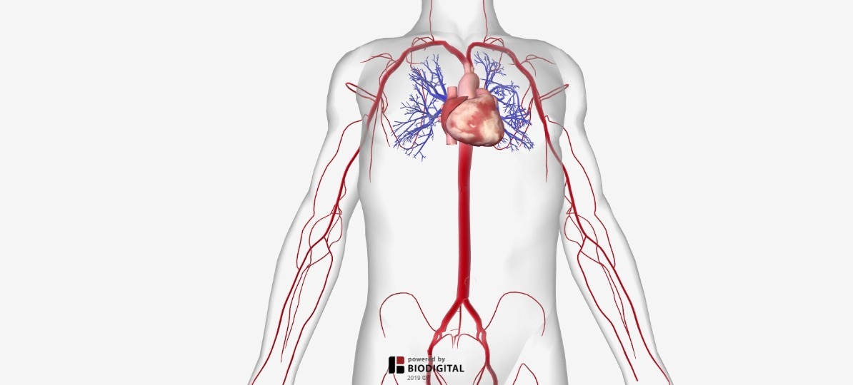


Arteries Of The Body Picture Anatomy Definition More
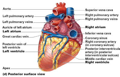


Coronary Sinus Aneurysm Incidental Discovery During Coronary Artery Bypass Grafting
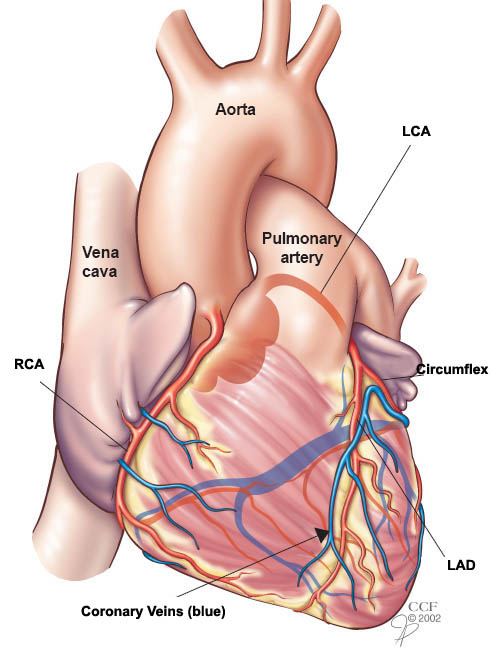
Heart and Vascular Coronary arteries supply blood to the heart muscle Like all other tissues in the body, the heart muscle needs oxygenrich blood to function Also, oxygendepleted blood must be carried away The coronary arteries wrap around the outside of the heart Small branches dive into the heart muscle to bring it blood Heart Bypass Surgery is an openheart surgery that is used to treat blockages of the heart arteries When there is a heart artery blockage, blood supply to areas of the heart are affected A heart bypass is attached beyond the blockage restoring blood flow to that area Heart bypasses are either arteries or veins taken from other parts of the> heart muscle contracting What sort of thing is expanding when you feel the pulse?


Interior View Of Human Chest Heart Lungs Arteries Veins Anatomy Stock Photo Download Image Now Istock
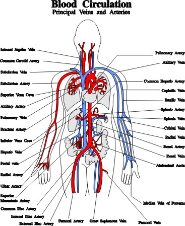


Blood Vessels Arteries Capillaries Veins Vena Cava Central Veins Lhsc
There are three arteries that run over the surface of the heart and supply it with blood (see the diagram above) There is one artery on the right side and two arteries on the left side of the heart The one on the right is known as the right coronaryThe base of the heart is located at the level of the third costal cartilage as seen in figure 1 There are typically Head and neck arteries The largest and most important artery in the circulatory system is the aorta The different parts of heart in heart diagram are discussed below Arteries and veins anatomy of the heartAnd coronary arteries are attached to the heart and transfer oxygenrich blood to your heart muscles Veins Although they are quite


1


Pedi Cardiology Anatomy Coronary Veins Coronary Arteries Arteries And Veins Coronary Arteries Arteries Anatomy
The right superior pulmonary vein passes in front of and a tad below the pulmonary artery at the root of the lung, and the inferior pulmonary vein is situated at the lowest part of the lung hilum In reference to the heart, the right pulmonary veins pass behind the right atrium and superior vena cava return, and the left pulmonary veins pass in labelled diagram of arteries Conclusion ArteriesCoronary circulation is the circulation of blood in the blood vessels that supply the heart muscle (myocardium) Coronary arteries supply oxygenated blood to the heart muscle Cardiac veins then drain away the blood after it has been deoxygenated Because the rest of the body, and most especially the brain, needs a steady supply of oxygenated blood that is free of all but theThe heart is an organ about the size of your fist that pumps blood through your body It is made up of multiple layers of tissue Your heart is at the center of your circulatory system This system is a network of blood vessels, such as arteries, veins, and capillaries, that carries blood to and from all areas of your body
:max_bytes(150000):strip_icc()/heart-and-circulatory-system-with-blood-vessels--97537745-a3bc2b2a6ca94390bfdf2696ad9bbddd.jpg)


Pulmonary Vein Anatomy Function And Significance


1
When the coronary arteries narrow to the point that blood flow to the heart muscle is limited (coronary artery disease), collateral vessels may enlarge and become active This allows blood to flow around the blocked artery to another artery nearby or to the same artery past the blockage, protecting the heart tissue from injury Coronary arteries and cardiac veins The heart is a muscular, fourchambered organ that is responsible for distributing blood throughout the body The continuous activity of the heart creates a large demand for nutrients to be delivered to cardiac tissue and for waste to be removed However, because the organ is several layers thick, it is not feasible for the tissue to Capillaries are small blood vessels between the arteries and the veins that circulate oxygenrich blood to the tissuesThey are extremely small and usually not even visible with the normal eye Veins drain deoxygenated blood from various parts of the body to the heart The pulmonary veins, however, bring oxygenated blood from the lungs to the heart
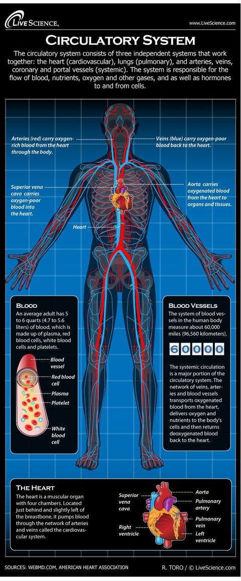


Human Circulatory System Diagram How It Works Live Science
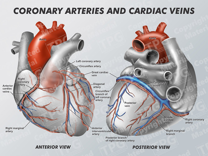


Coronary Arteries And Cardiac Veins Order
Diagram Blood flow through the right side of the heart involving the following cardiac structures superior vena cava (SVC), inferior vena cava (IVC), right atrium (RA), tricuspid valve (TV), right ventricle (RV), pulmonary valve (PV), and main pulmonary artery (PA)Arteries, veins, and the heart are the main parts of the system To understand the system, the students need to create their diagrams It can also help them in getting an overview of artery vs vein Creating a freehand diagram of arteries and veins can be troublesome The students can use online tools like EdrawMax, which can help them create Arteries Arteries are blood vessels that transport oxygenated blood from the heart to various parts of the body They are thick, elastic and are divided into a small network of blood vessels called capillaries The only exception to this is the pulmonary arteries, which carries deoxygenated blood to the lungs Veins


14 4 Blood Vessels Human Biology
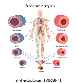


Arteries Veins Capillaries Diagram High Res Stock Images Shutterstock
Heart Arteries and Veins Diagram Quizlet Upgrade to remove ads Only $299/monthThe arteries are the blood vessels that deliver oxygenrich blood from the heart to the tissues of the body Each artery is a muscular tube lined by smooth tissue and has three layers The intima_____ 2 Blood vessels that carry blood away from the heart are called _____ 3
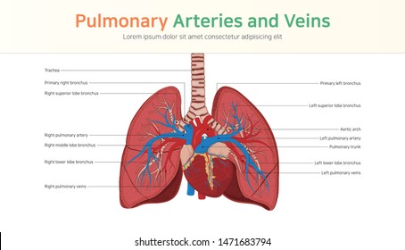


Pulmonary Veins Images Stock Photos Vectors Shutterstock
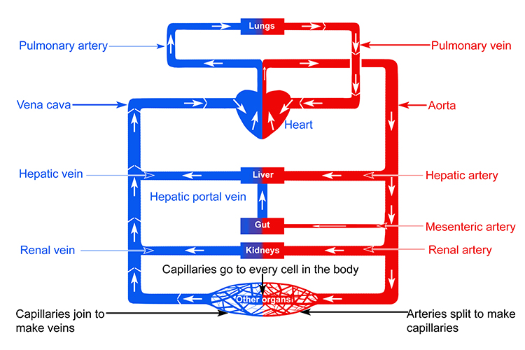


Labelled Diagram Of Vein Valves And Vein Artery Circuit
Major arteries By definition, an artery is a vessel that conducts blood from the heart to the periphery All arteries carry oxygenated blood–except for the pulmonary arteryThe largest artery in the body is the aorta and it is divided into four parts ascending aorta, aortic arch, thoracic aorta, and abdominal aorta After receiving blood directly from the left ventricle of the heart, theUpper Body Circulation In the lungs, the pulmonary arteries (in blue) carry unoxygenated blood from the heart into the lungs Throughout the body, the arteries (in red) deliver oxygenated blood and nutrients to all of the body's tissues, and the veins (in blue) return oxygenpoor blood back to the heart The aorta is the large artery leaving the heart The superior vena cava is the largeAlthough it is a single organ, the heart functions as a double pump, with arteries carrying blood away from and veins carrying blood toward the heart The right side works as the pulmonary circuit pump It receives oxygenpoor blood from the veins of


Anatomy Of The Coronary Circulation Angiographic Visualization Dr


Coronary Veins Cardiac Veins
Look at Figure B It shows arteries and veins within the human body Each artery and vein branches out to tiny capillaries Write the correct term in each blank to answer the questions or complete the sentence 1 What pumps blood through your body? 14 Heart Arteries Diagram Labeled Labeled heart diagram showing the heart from anterior Inner body parts with their names CoronaryArteriesComplete from facultyetsuedu (taken from johnson, weipz and savage lab book) This is an excellent human heart diagram which uses different colors to show different parts and also labels a numberCoronary arteries are located on top of the heart and supply the heart itself with blood 2 The first visible branch from the aorta is the brachiocephalic artery , it divides into the right common carotid artery , which supplies the right side of the neck, and the right subclavian artery , which supplies the right shoulder and arms


What Is The Difference Between An Artery And A Vein


Coronary Artery And Vein Anatomy Model
The network of veins, arteries and blood vessels transports oxygenated blood from the heart, delivers oxygen and nutrients to the body's cells and then returns deoxygenated blood back to the heart Pulmonary artery is the only artery that takes deoxygenated blood from the right side of your heart to your lungs;Features of Arteries and Veins (With Diagram) In this article we will discuss about the general features of Arteries and Veins Arteries and veins are main blood vessels Arteries carry blood from the heart to different body parts Veins bring blood from different body parts to the heart The veins have valves to prevent backward flow of blood


Www Pearsonhighered Com Assets Samplechapter 0 1 3 4 Pdf


What S The Difference Between Veins And Arteries Britannica
Start studying heart arteries and veins Learn vocabulary, terms, and more with flashcards, games, and other study tools


Coronary Veins Radiology Reference Article Radiopaedia Org
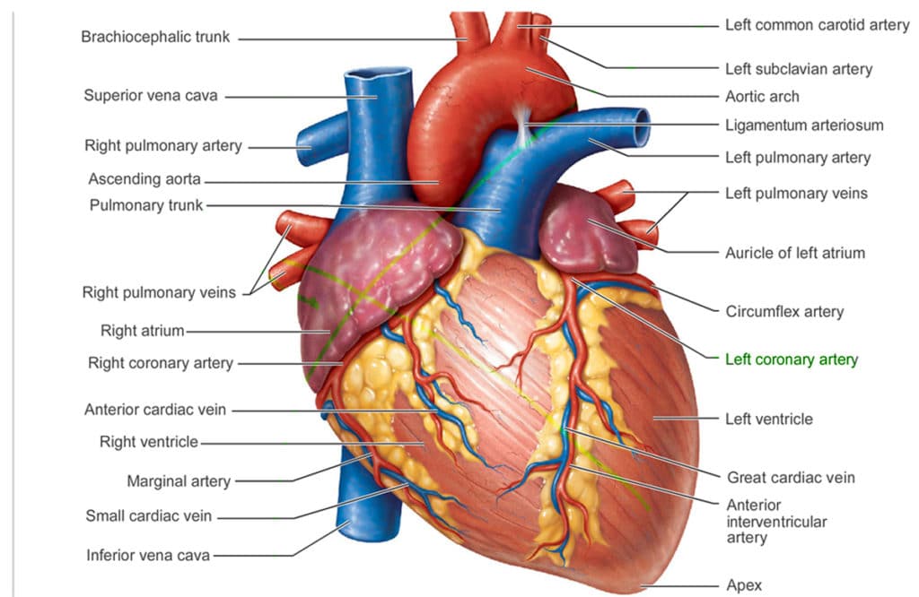


The Anatomy Of A Heart Central Georgia Heart Center
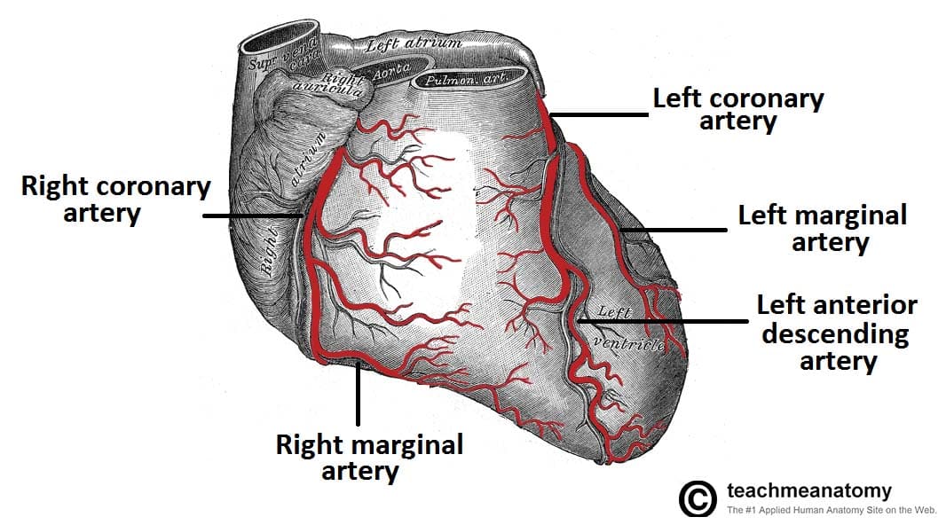


Vasculature Of The Heart Teachmeanatomy


Coronary Artery Anatomy
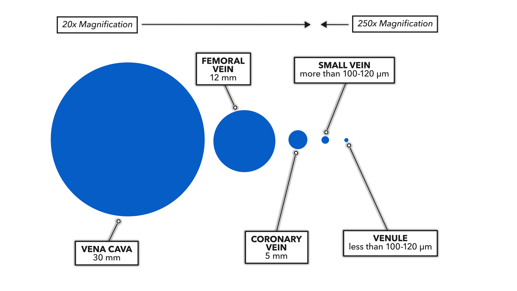


Crossfit The Heart Part 6 Blood Vessel Basics
:watermark(/images/watermark_only.png,0,0,0):watermark(/images/logo_url.png,-10,-10,0):format(jpeg)/images/anatomy_term/sinus-coronarius-3/BTxnNt3FhCAKDchLsEFDA_Sinus_coronarius_01.png)


Coronary Arteries And Cardiac Veins Anatomy And Branches Kenhub


Cardiac Anatomy Thoracic Key


Coronary System Tutorial What Is The Coronary System


Anatomy Of The Human Heart Physiopedia


Cardiac Veins An Anatomical Review Sciencedirect


Blood Vessels Circulatory Anatomy


Arteries And Veins Png Images Pngegg


Pedi Cardiology Anatomy Coronary Veins Coronary Arteries Coronary Arteries Coronary Arteries


Labelled Diagram Of Vein Valves And Vein Artery Circuit


Heart Anatomy Labeled Diagram Structures Blood Flow Function Of Cardiac System Ezmed


Heart Knowledge Amboss


Coronary Veins Comprehensive Ct Anatomic Classification And Review Of Variants And Clinical Implications Radiographics


Structure Of The Heart Biology For Majors Ii


Cardiovascular Media Library Watch Learn Live


Pin On Bio 12 Circulatory Project


Coronary Artery Disease Causes Symptoms Diagnosis Treatments


5 Heart Supply With Overview Of Arteries And Veins Download Scientific Diagram


Vascular System 1 Anatomy And Physiology Nursing Times
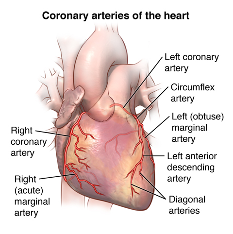


Anatomy And Function Of The Coronary Arteries Johns Hopkins Medicine


The Normal Heart
/heart-and-circulatory-system-with-blood-vessels--97537745-a3bc2b2a6ca94390bfdf2696ad9bbddd.jpg)


Pulmonary Vein Anatomy Function And Significance


Istaroxime Clinical Trials Arena
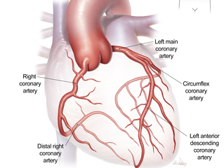


Cardiovascular System


Cardiac Arteries Veins Anterior Diagram Quizlet


Torso With The Heart Artery Vein And Capillary Labeled Media Asset Niddk


Blood Vessel Definition Anatomy Function Types Britannica
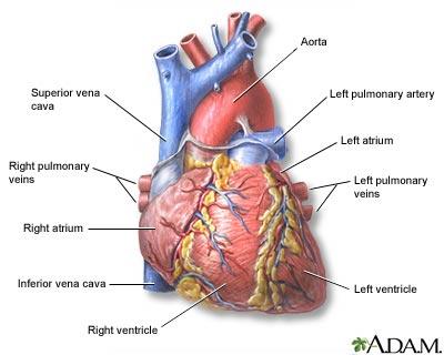


Heart Front View Medlineplus Medical Encyclopedia Image


Arteries Veins Of The Heart Diagram Quizlet
:watermark(/images/watermark_only.png,0,0,0):watermark(/images/logo_url.png,-10,-10,0):format(jpeg)/images/anatomy_term/vena-cardiaca-magna/7egsYCVRb0d6wMelbewyQ_V._cardiaca_magna_01.png)


Coronary Arteries And Cardiac Veins Anatomy And Branches Kenhub
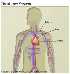


Anatomy And Circulation Of The Heart


Coronary Circulation Wikipedia


The Principal Arteries And Veins
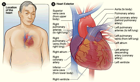


3 Anatomy Of The Coronary Arteries Atrain Education


27 Differences Between Arteries And Veins Arteries Vs Veins
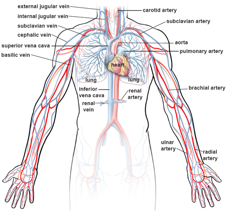


Illustrations Of The Blood Vessels
/vascular-system-veins-56c87fa03df78cfb378b3e7c.jpg)


What Is A Vein Definition Types And Illustration


Great Cardiac Vein Radiology Reference Article Radiopaedia Org
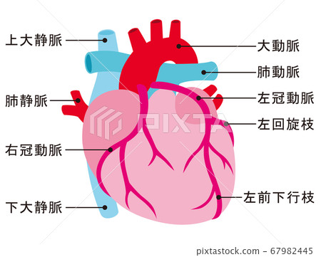


Heart Artery Vein Coronary Artery Stock Illustration
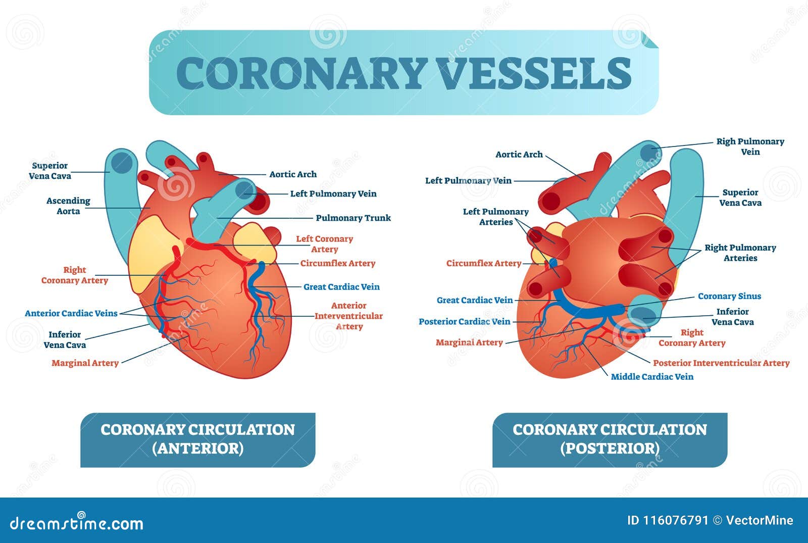


Coronary Vessels Anatomical Health Care Vector Illustration Labeled Diagram Heart Blood Flow System With Blood Vessel Scheme Stock Vector Illustration Of Heartbeat Cardio
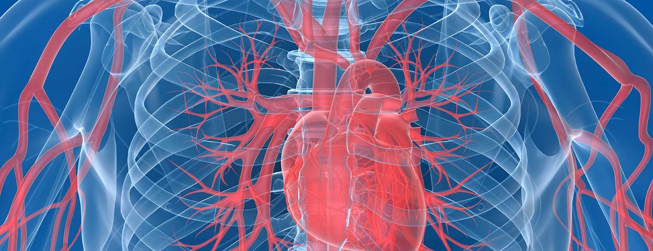


Anatomy And Function Of The Coronary Arteries Johns Hopkins Medicine


Blood Vessels Educational Banner Or Poster Comparison Of The Structure Of The Artery And Vein Diagram For The Study Of Canstock
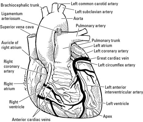


Arteries And Veins That Feed The Heart Dummies
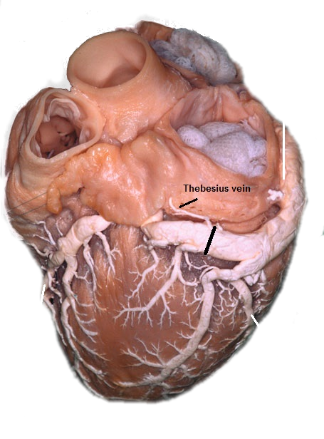


Cardiac Veins Cthsurgery Com
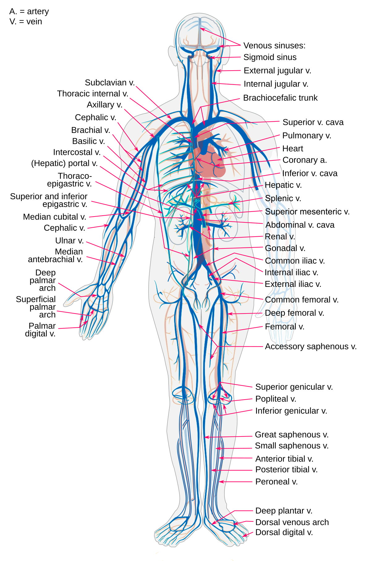


Vein Wikipedia


Coronary Sinus And Great Cardiac Vein Anatomy Posterior Surface View Of Heart
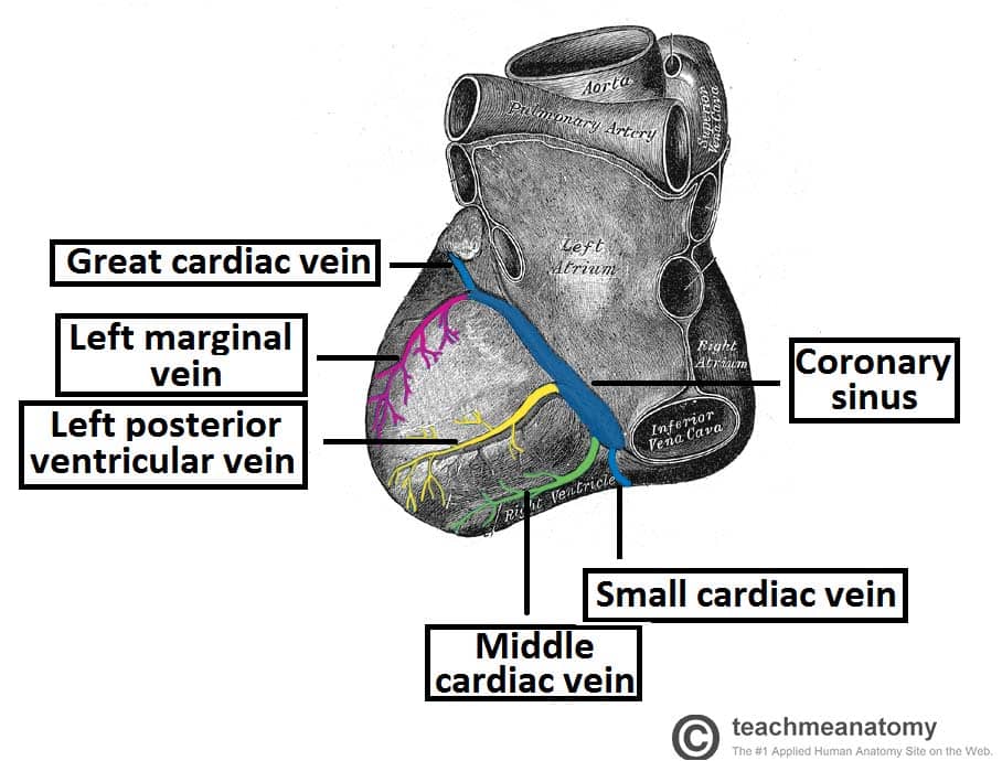


Vasculature Of The Heart Teachmeanatomy
:watermark(/images/watermark_5000_10percent.png,0,0,0):watermark(/images/logo_url.png,-10,-10,0):format(jpeg)/images/overview_image/265/eLfoZ6uTZlVZPyfTksFfgw_coronary-arteries-and-cardiac-veins_english.jpg)


Coronary Arteries And Cardiac Veins Anatomy And Branches Kenhub


Clinical Anatomy Cardiac Coronary Vessels Left And Right Coronary Artery Venous Sinus Youtube


Anatomy Of The Heart And Major Coronary Vessels In Anterior Left And Download Scientific Diagram


Coronary Circulation Of The Heart Bioscience Notes


Your Heart Blood Vessels


Coronary Arteries Anatomy Blood Supply Of Heart Arterial Supply Of Heart Animation Youtube


Artery And Vein Diagram For Powerpoint Pslides
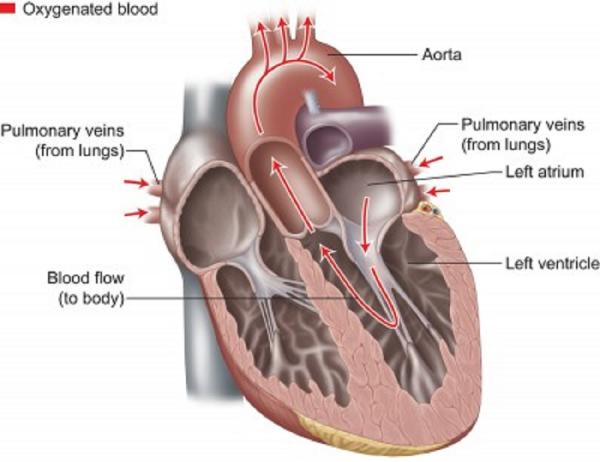


How The Heart Blood Vessels Work Heart Vascular Institute Temple Health
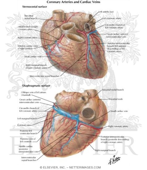


Arteries And Veins Of The Heart Coronary Arteries And Cardiac Veins


1
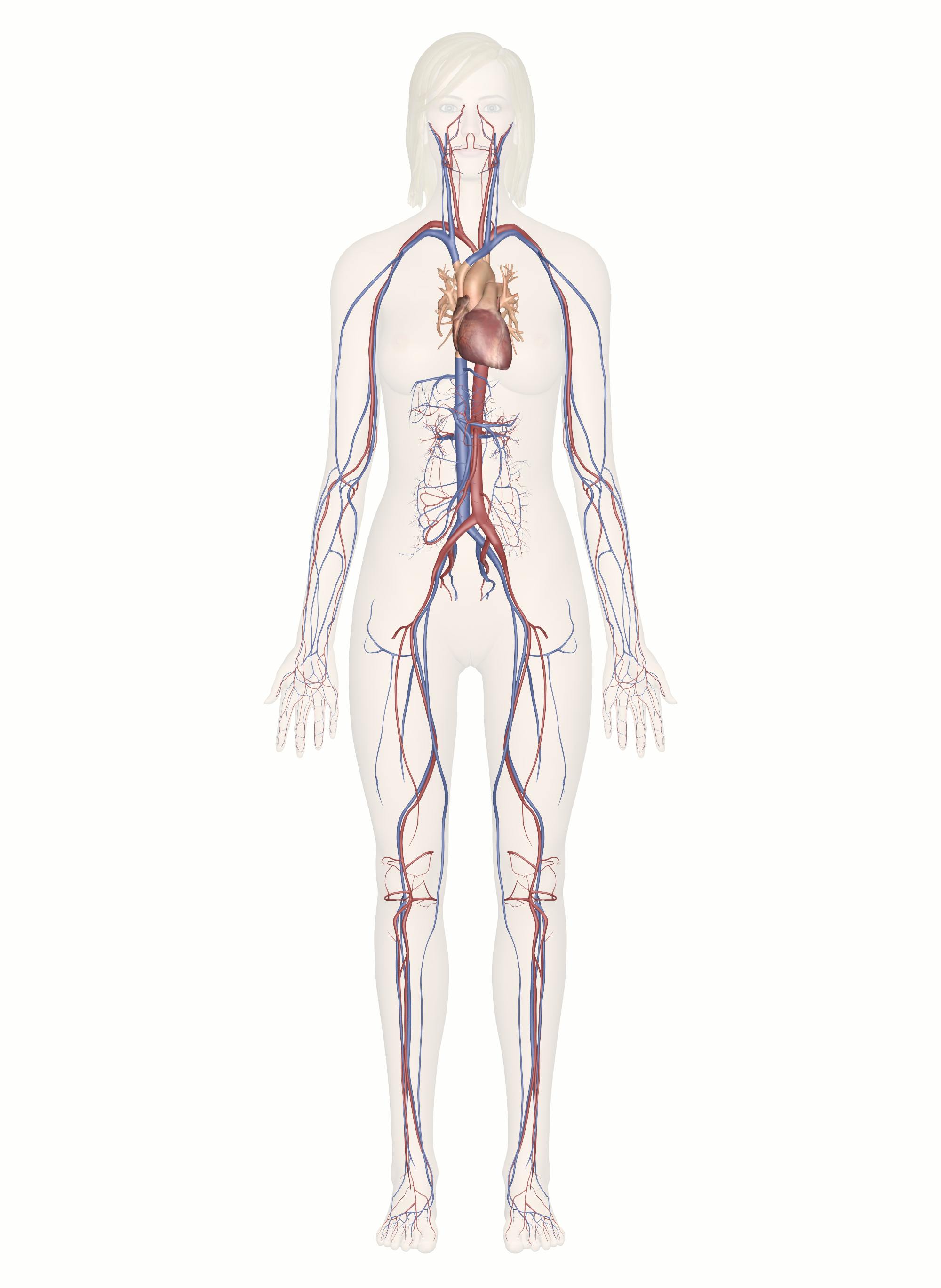


Cardiovascular System Human Veins Arteries Heart
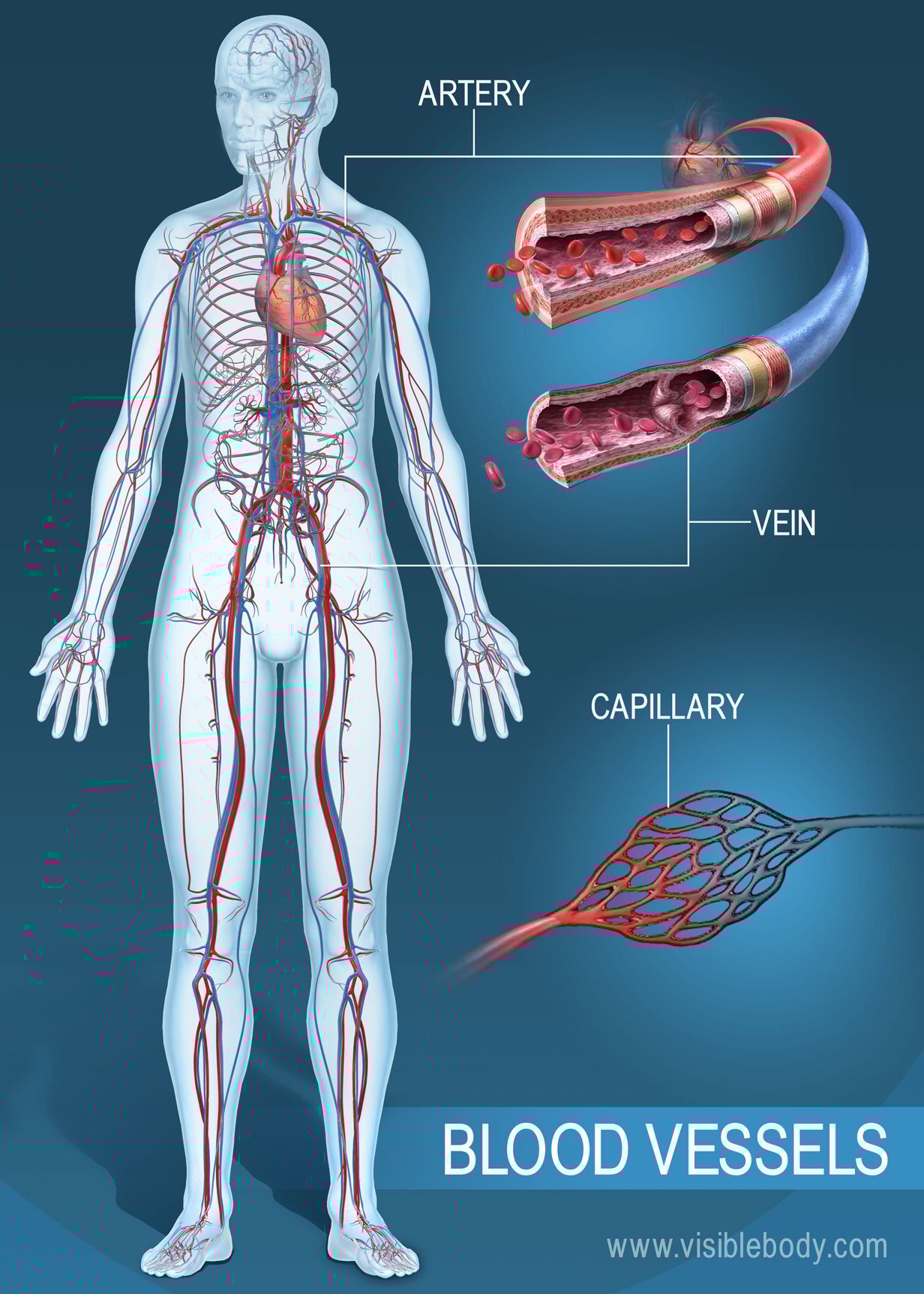


Blood Vessels Circulatory Anatomy


40 3b Arteries Veins And Capillaries Biology Libretexts
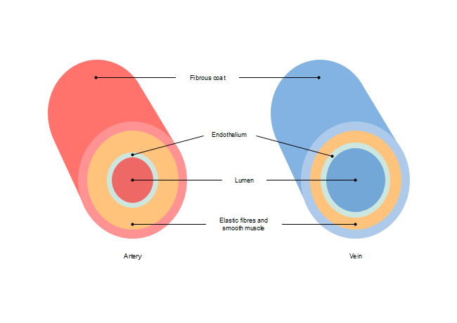


Free Artery Vein Diagram Templates


19 The Coronary Veins Radiology Key


Mammalian Heart And Blood Vessels Boundless Biology


1


Cardiac Veins An Anatomical Review Sciencedirect


How The Heart Works Diagram Anatomy Blood Flow


Interior View Of Human Chest Heart Lungs Arteries Veins Anatomy Stock Photo Download Image Now Istock
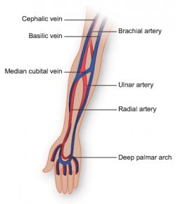


Vasculature Of The Arm Texas Heart Institute


Arteries Of The Body Picture Anatomy Definition More
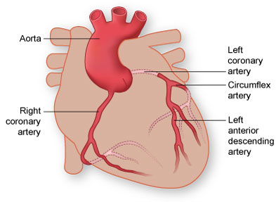


Coronary Arteries Texas Heart Institute


Artery Wikipedia
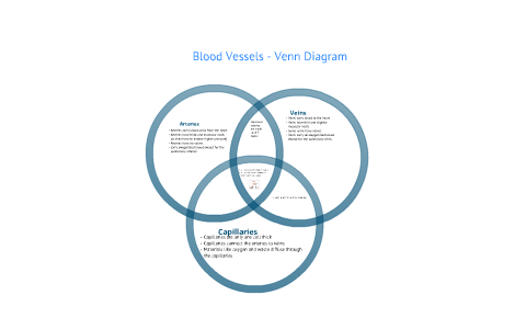


Blood Vessels Venn Diagram By David Berry
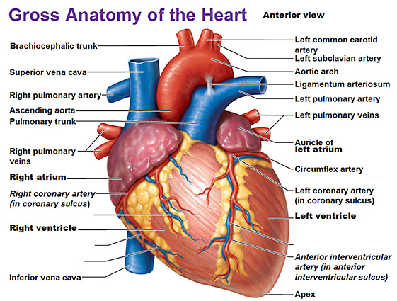


Heart Anatomy
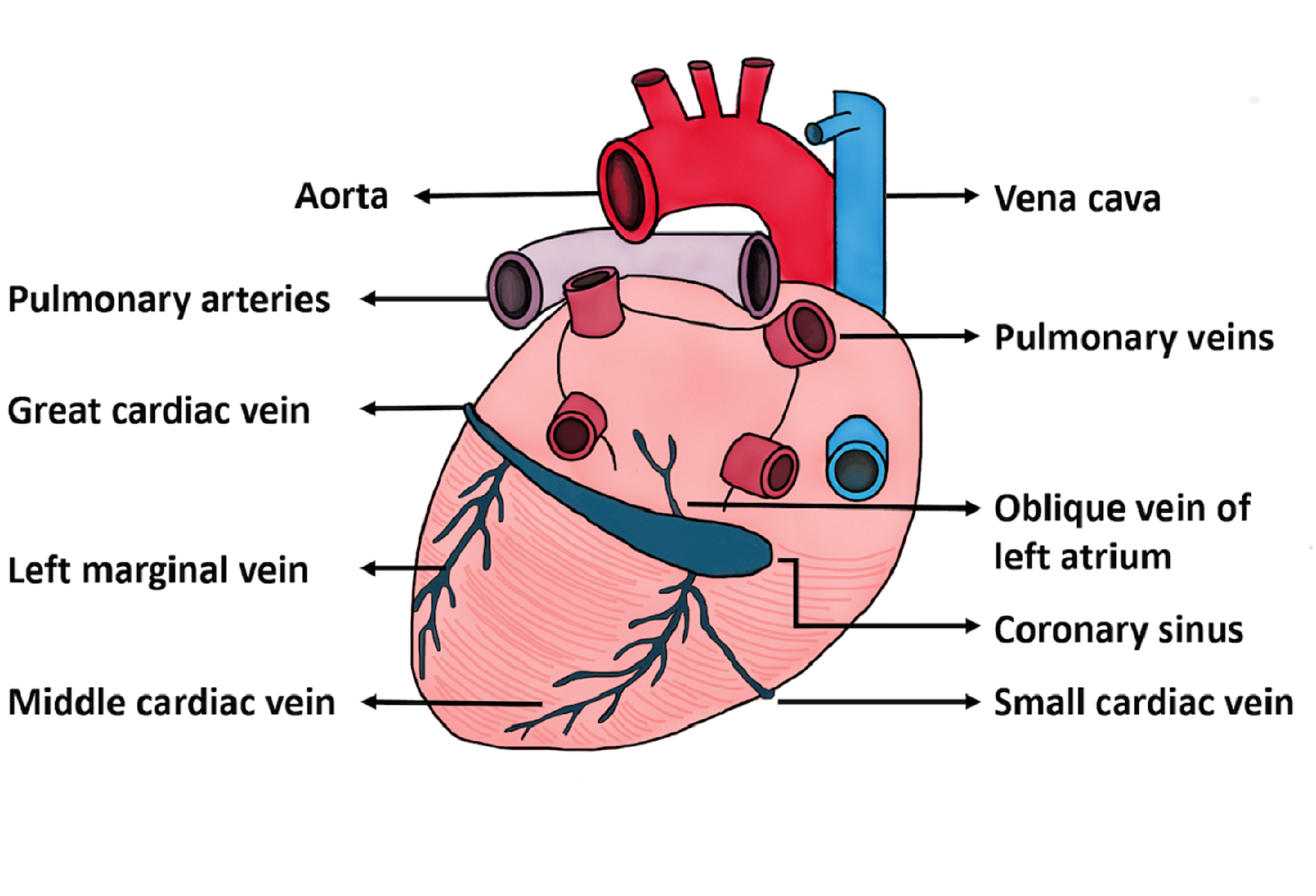


Cureus A Route Less Traveled Anomalous Venous Drainage Of The Right Heart


0 件のコメント:
コメントを投稿